Abdelaaty A Shahat1,2 ![]() ,
Abeer Y Ibrahim3,
Amal AM Al-Ghamdi4,
Mansour S Alsaid1
,
Abeer Y Ibrahim3,
Amal AM Al-Ghamdi4,
Mansour S Alsaid1
For correspondence:- Abdelaaty Shahat Email: aashahat@hotmail.com Tel:+966537507320
Received: 19 March 2016 Accepted: 17 July 2016 Published: 30 August 2016
Citation: Shahat AA, Ibrahim AY, Al-Ghamdi AA, Alsaid MS. Phytochemical investigation of Rhus tripartita and its activity against cyclooxygenases and acetylcholinesterase. Trop J Pharm Res 2016; 15(8):1697-1706 doi: 10.4314/tjpr.v15i8.15
© 2016 The authors.
This is an Open Access article that uses a funding model which does not charge readers or their institutions for access and distributed under the terms of the Creative Commons Attribution License (http://creativecommons.org/licenses/by/4.0) and the Budapest Open Access Initiative (http://www.budapestopenaccessinitiative.org/read), which permit unrestricted use, distribution, and reproduction in any medium, provided the original work is properly credited..
Purpose: To investigate the inhibitory activity of crude methanol extract, fractions and two pure compounds from Rhus tripartita stem cyclooxygenases (COX-1 and COX-2) and acetylcholinesterase (AChE); also, to evaluate their antioxidant properties in in-vitro assays.
Methods: R. tripartita extract and fractions were examined as inhibitors of COX-1 and COX-2 and AChE. Their antioxidant properties were also evaluated using various antioxidant tests, including free radical scavenging, nitric oxide, and total antioxidant capacity.
Results: Ethyl acetate fraction (RT2), aqueous fraction (RT4), subfraction RT2-II, and subfraction RT2-III had a potent inhibitory effect on AChE with low IC50 values. At the same time, a subfraction (RT2-III) was an effective COX-2 inhibitor (94.5 % at 10 µg/mL) followed by ethyl acetate fraction (RT2; 79.39 %), aqueous fraction (RT4; 70 %), total extract (RT; 66.18 %), subfraction RT2-III (64.08 %), compound 5 (epicatechin; 61.25 %), compound 2 (taxifolin; 55.63 %), subfraction RT2-II (46.85 %), and butanol fraction (RT3; 40.80%). RT2, RT4, RT2-II, RT2-III, and compound 2 each showed 100 % inhibition at 20 µg/mL.
Conclusion: The ethyl acetate fraction (RT2), aqueous fraction (RT4) and subfractions RT2-II and RT2-III are effective natural inhibitors of AChE. Subfraction RT2-III is the best COX-2 inhibitor, followed by ethyl acetate fraction RT2 and aqueous fraction (RT4). These bioactive materials can be considered natural COX-2 and AChE inhibitors.
Introduction
The search for innovative pharmacologically active natural agents for dealing with human diseases can lead to the discovery of several clinically valuable natural drugs. Many diseases like rheumatoid arthritis, chronic asthma, multiples sclerosis, inflammatory bowel disease, psoriasis and neuro-inflammatory diseases are related to inflammation [1]. Inflammation is a mandatory host defense reaction to infection or injury [2]. It is accepted that both the beneficial and detrimental effects of nonsteroid anti-inflammatory drugs (NSAIDs) are attributable to their ability to inhibit prostaglandin synthesis through direct blockage of cyclooxygenase 2 (COX-2). Of the two isoforms of COX, COX-1 is a constitutively expressed form required for common physiological functions, and COX-2 is induced only as a part of inflammatory processes [3]. Therefore, a selective COX-2 inhibitor can avoid toxic effects on cellular physiological functions. The cascades of an inflammatory response are known to be the main risk factor of neurodegenerative diseases including AD, which is a devastating disorder manifesting itself in deterioration of memory and cognition, impairments in execution of activities of daily living, and many behavioral and neuropsychiatric aberrations [4]. Rhus species are widely used in both modern and traditional medicine due to their high concentration of polyphenols, flavonoids, proanthocyanidins, and other phytochemicals. A Rhus extract was reported to serve as an antimicrobial, antitumorigenic, antioxidant, hypoglycemic, and anticonvulsant agent [5]. Recent studies on Rhus tripartita confirmed the abundant presence of proanthocyanidins in a root cortex extract [6]. Proanthocyanidins isolated from different plants were reported as potent antioxidant, antiviral, and anti-HIV agents [7].
The present study was designed to evaluate an R. tripartita extract, its fractions, and pure compounds as anti-inflammatory agents acting against COX-1 and COX-2, and to test them on acetylcholinesterase activity in addition to evaluation of their antioxidant properties.
Methods
Plant material
Rhus tripartita (Ucria) Grande (Anacardiaceae) was collected from Hail area, Kingdom of Saudi Arabia in April 2013. Hail is located in the North-Western of Kingdom of Saudi Arabia at latitude 27º31' North and longitude 41º43' East. The temperature is between 9.6-33.0 ºC and an annual rainfall of about 29.4 mm. The average relative humidity is 90-100%. Moreover, elevation heights above sea level about 1009.1 m. The plant was identified by Dr. Mohamed Yousuf. A voucher specimen (SY202/2013) was deposited in the Herbarium of the College of Pharmacy, King Saud University, Riyadh, Kingdom of Saudi
Reagents
Peroxidase, 2,2-azino-bis(3-ethylbenzthiazoline-6-sulfonic) acid diammonium salt, phenazine methosulphate (PMS)-nicotinamide adenine dinucleotide (NADH), nitroblue tetrazolium (NBT), Tris-HCl buffer, Greiss reagent, sodium nitroprusside ascorbic acid (Vc), butylated hydroxytoluene (BHT), leuco-2,7-dichlorofluorescein diacetate, hematin, arachidonic acid, Tris-buffer, cyclooxygenase enzyme (COX-1 from sheep, EC 1.14.99.1 or COX-2), acetylcholinesterase (EC No. 3.1.1.7) type VI-S (from electric eel), eserine hemisulfate salt, acetylthiocholine iodide, and dithiobis (nitrobenzoic) acid (DTNB) were purchased from Sigma-Aldrich, USA
Extraction of plant materials and isolation of pure compounds
Air-dried powdered stem (1000 g) were exhaustively extracted with 80 % methanol (3 x 3000 mL) at room temperature. After filtration and evaporation using reduced pressure at 35 °C, the residual aqueous solution (RT) was partitioned with methylene chloride (CH2Cl2; fraction RT1), ethyl acetate (EtOAc; fraction RT2), or normal butanol (n-BuOH; fraction RT3) (4 400.mL each). The organic fractions were concentrated using reduced pressure, and the aqueous residual layer was lyophilized (RT4). The EtOAc fraction (RT2; 20 g) was separated on a Sephadex LH-20 column (Pharmacia; 100 x 4 cm) using EtOH as the mobile phase. Fractions 50 mL each were collected and analyzed by thin-layer chromatography (TLC; Silica gel 60 F254, 0.2 mm, Merck) using the upper layer of the mixture EtOAc-HOAc-HCOOH-H2O (30:0.8:1.2:8) as a mobile phase.
The spots were detected by UV light (254 and 366 nm) before and after spraying with Neu’s spray reagent (reagent a) and vanillin H2SO4 (reagent b). Fractions 20-35, which gave positive reaction with reagent a and developed a purple-red color with reagent b, were combined (RT2-I). Fractions 36-43 which gave a yellow or orange color with reagent a, were combined in one group (RT2- II) and Fraction 44-69, fraction 70-85 and 86-102 which developed a purple-red color with reagent b, were collected together in one group (RT2- III), (RT2- IV) and (RT2- VI).
Column chromatography of sub-fractions RT2-I on Sephadex LH-20 using methanol as mobile phase, gave compounds 1 and 2. Sub-fraction RT2-II was subjected to Sephadex LH-20 using methanol as mobile phase to give compounds 3. Sub-fraction R2-III was subjected to C18-reverse-phase columns with MeOH-H2O (6:4) as eluent to give compounds 4 and 5. Sub fraction (R2- IV), (R2- V) and (R2- VI) are kept for further investigation.
Determination of inhibitory activity on cyclooxygenases
The method of Larsen et al [8] was used for testing of natural or synthetic materials against cyclooxygenase 2. The main idea behind this method is oxidation of leuco-2,7-dichlorofluorescein in the presence of phenol to produce hydrogen peroxide. This method is considered a sensitive spectrophotometric assay for the PGH synthesis activity. Leuco-2, 7-dichlorofluorescein di acetate (L-DCF) was hydrolyzed in sodium hydroxide for 10 min and then the alkaline media was neutralized using hydrochloric acid and finally the reaction micture was diluted with Tris-buffer. The tested enzyme (COX-1 or COX-2) was diluted in Tris-buffer in order that a known aliquot gave an absorbance change of 0.05/min in the test reaction. The diluted enzyme was incubated with samples or equivalent volume of methanol in presence of hematin. L-DCF, arachidonic acid and phenol were added to enzyme mixture to begin the reaction. The reaction was recorded spectrophotometrically (Jasco, serial No. C317961148, Japan) over 1 min at 502 nm. A blank reaction mixture was recorded for each sample to avoid any non-enzymatic activity attributed to the test sample and another reaction for reference compound, Celecoxib, was also monitored at the same time at the same concentration of samples.
Acetylcholinesterase inhibition determination
Enzymatic activity was analyzed using an adaptation of the method of Ingkaninan [9]. Namely, 500 µL of the DTNB "3 mM", 100 µL of acetylthiocholine iodide (15 mM), 275 µL of Tris–HCl buffer (50 mM, pH 8), and 100 µL of each sample at different concentrations (2.5, 5, 10, 20, or 40 µg/mL) was dissolved in ethyl alcohol (HPLC grade) and was added to a 1-mL cuvette then was used as a blank. In the reaction cuvette, 25 µL of Tris-HCl buffer was replaced by the same volume of an enzyme solution containing 0.28 U mL−1. The color developed was monitored for 5 min at 405 nm. The percent inhibition was calculated using the formula: {(control absorbance − sample absorbance)/control absorbance} x 100. The IC50 was calculated by log-probit analysis. Eserine served as a positive control [10].
Nitric oxide radical scavenging activity assay
The test samples at 2.5, 5, 10, 20, and 40 µg/mL were tested as a nitric oxide radical scavenger using a sodium nitroprusside (SNP) system generating NO•. Nitric oxide radicals (NO•) were generated from SNP in the aqueous solution at physiological pH, and this radical reacts with oxygen to produce nitrite ions which were detected by the Greiss reagent [11]. Two milliliters of the above reaction mixture containing tested fractions or standard compounds and SNP (final concentration, 10 mM) in phosphate buffered saline (pH 7.4) were incubated at 25 °C for 150 min. One milliliter of the Greiss reagent was added to 1 mL of the incubated mixture, and then absorbance was measured at 540 nm against a blank solution. The blank solution contained all chemicals and reagents without samples and was incubated for the same period.
Free radical scavenging activity assay
Free radical scavenging activity was measured using method of Yamaguchi et al [12]. In brief 0.1 mM of DPPH solution was added to 3 mL of each sample or standards at 2.5, 5, 10, 20 and 40 µgmL-1. The reaction mixture was shaken and allowed to stand at room temperature for 30 min. The absorbance was measured at 517 nm. DPPH• radical scavenging activity (D) was calculated in the reaction medium as in Eq 1.
D (%) = 100 – {(A0 - A1)/A0)}100 …………. (1)
A0 is the absorbance of the control reaction, and A1 is the absorbance in the presence of the sample.
Superoxide anion-scavenging activity
This activity was isolated by the method of Gülçin et al [13], where superoxide radicals are generated in a phenazine methosulphate (PMS)–nicotinamide adenine dinucleotide (NADH) system via oxidation of NADH; the superoxide radicals are assayed by the reduction of NBT. Superoxide radicals were generated in Tris-HCl buffer (16 mM, pH 8.0, 3 mL) containing NBT (50 µM, 1 mL), NADH (78 µM, 1 mL), and fractions or isolated compounds to be assayed (1 mL) at different concentrations. The reaction was started by addition of a phenazine methosulfate (PMS) solution (10 µM, 1 mL). The reaction mixture was incubated at 25 °C for 5 min, and absorbance (560 nm) was measured. Decreasing absorbance indicated increasing superoxide anion-scavenging activity. The percentage inhibition of superoxide anion production was calculated using Eq 2:
Inhibition (%) = {(A0 - A1)/A0}100 …………. (2)
where A0 is the absorbance of the control, and A1 is the absorbance of the tested materials.
Total antioxidant activity
This capacity of the tested materials was measured using an 2,2-azino-bis(3-ethylbenzthiazoline-6-sulfonic acid) (ABTS) peroxidase system as explained by Miller et al. [14]. Peroxidase (4.4 U/mL) was kept in the dark with hydrogen peroxide (50 μM) and ABTS (diammonium salt, 100 μM) for 60 min, and the absorbance of the developed color was determined for each sample, standard, and control at 734 nm. Total antioxidant capacity (C) was then calculated as in Eq 3.
C (%) = {1 - (As/Ac)}100 ……………..... (3)
where As and Ac are the absorbance of test sample and control, respectively.
Statistical analysis
All data are expressed as mean ± standard deviation (SD). One-way analysis of variance was used for statistical evaluation of significant differences among multiple groups followed by a post hoc test: Tukey’s multiple-comparison test. Differences were considered significant at p < 0.05. The analysis was performed with SPSS 9 software.
Results
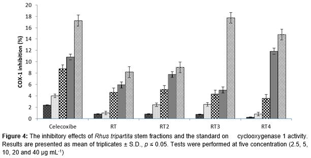
The crude methanol extract, different fractions, and two pure compounds as well as the standard compound, celecoxib, were screened for their possible inhibitory effect on cyclooxygenases (COX-1 and COX-2). All of the tested samples inhibited COX-2 in a concentration-dependent manner reaching 100 % in efficiency at the maximum concentration (40 µg/mL), whereas celecoxib reached 29.36 % efficiency at the same concentration (Figs. 1 and 2). Subfraction RT2-III showed the best COX-2 inhibition: 94.5 % inhibition at 10 µg/mL, followed by fraction RT2 (79.39 %), fraction RT4 (70%), total extract RT (66.18 %), subfraction RT2-I (64.08 %), compound 5 (61.25 %), compound 2 (55.63 %), subfraction RT2-II (46.85 %), and then fraction RT3 (40.80 %). Fraction RT2, fraction RT4, subfraction RT2-II, subfraction RT2-III, and compound 2 each showed 100 % inhibition at 20 µg/mL (Figs. 1 and 2).
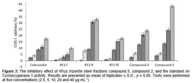
On the other hand, the nine tested fractions showed weak inhibitory effect against cyclooxygenase-1. The inhibitory effect of leuco-dichlorofluorescein (L-DCF) oxidation in the presence of phenol by the hydroperoxide formed in the COX-1 reaction increased with the increasing concentration of a tested fraction. The maximal inhibitory effect on COX-1 was produced by compound 5 (43.34 %) followed by Rt2-II (32.73 %), and compound 2 (23.81 %), while fractions RT2-III showed the same inhibitory effect as that of celecoxib (17 %; ). The oxidation was only weakly suppressed by fractions RT (8.17 %), RT2 (9.01 %), or Rt2-I.
The Rhus fractions and isolated compounds were also tested for their probable inhibitory effects in the in-vitro assay, against AChE. They inhibited acetylcholinesterase concentration-dependently. Acetylcholinesterase significantly inhibits at 2.5 µg/mL when it was treated with Sub-fraction RT2-III (49.81 %) followed by fraction RT2 (42.56 %), RT-4 (41 %) and then Sub-fraction RT2-I (33.14 %) while compound 5 and compound 2 showed the weakest inhibitory effects (6.16 and 3.62 %, respectively Table, 1). Total extract (RT) and fraction RT3 showed the smallest suppressive effect toward acetylcholi-nesterase at 2.5 µg mL-1 (16.72 % and 15.44 %, respectively). On the other hand, the enzyme was completely inhibited (100 %) when it was treated with fractions RT2 and Sub-fraction RT2-III at 20 µg/mL, while the total extract RT, fraction RT2, fraction RT4 and Sub-fraction RT2-III at 40 µg/mL Sub-fraction RT2-III, fraction RT2, RT4 and Sub-fraction RT2-I were the potent inhibitor for acetylcholinesterase, they presented the lowest IC50 values (3.42, 4.10, 4.45 and 6.26 µg/ mL, respectively).
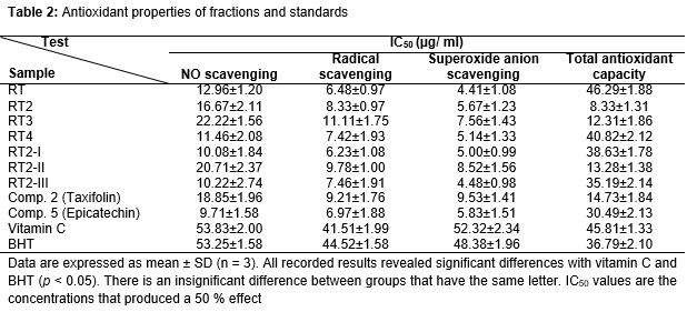
Antioxidant properties of natural substances play an important role in defense mechanism or treating many diseases. Antioxidant properties were assessed in in-vitro methods. The total extract, fractions, and compounds scavenged nitric oxide radicals, DPPH radicals and superoxide anion radicals and they were more potent than tested reference compounds, vitamin C and BHT. Compound 2, subfraction RT2-I, and subfractions RT2-III and RT4 have a valuable nitric oxide radical-scavenging effect with low IC50 values (9.71, 10.08, 10.22, 11.46, and 12.96 µg/mL, respectively), while the minimal recorded scavenging effect was shown by RT4. Fraction RT3 and subfraction RT2-II showed the second weakest scavenging action (22.22 and 20.71 µg/mL; ). On the other hand, DPPH radicals were significantly captured by R. tripartita fractions in the following order: subfraction RT2-I, total fraction RT, compound 5, fraction RT4, and subfraction RT2-III, with insignificant differences among them ().
Superoxide anion radicals were trapped by R. tripartita fractions, and the crude stem extract turned out to have the minimal IC50 of 4.41 µg/mL. Subfraction RT2-I, fractions RT4 and RT2, and compound 5 were alike in terms of quenching the SO2- radicals: they showed the same IC50 values with insignificant differences (IC50 ≈ 5 µg/mL). The long-lived ABTS•+ radical was strongly quenched by RT2 (IC50 = 8.33 µg/mL) while RT3, subfraction RT2-II, and compound 5 showed nearly the same effect (IC50 values were 12.31, 13.28, and 14.73 µg/mL, respectively). R. tripartita total extract had the same strength of the effect as vitamin C did, whereas subfraction RT2-III and compound 5 mimicked the effect of BHT (35.19 and 30.49 µg/mL, respectively).
NMR data of isolated compounds
Gallocatechin (1)
1H NMR (CD3OD): δ 6.41 (2 H, H-2', H-6'), 6.41 (2 H, H-2', H-6'), 5.93 and 5.87 (2 H, H-6, H-8), 4.55 (1 H, H-2), 4.53 (1 H, H-3), 2.84 (1 H, H-4a), 2.80 (1 H, H-4b).
13C NMR (CD3OD): δ 81.46 (C-2), 67.36 (C-3), 26.62 (C-4), 155.44 (C-5), 94.8 (C-6), 156.4 (C-7), 99.3 (C-8), 156.2 (C-9), 99.3 (C-10), 130.1 (C-1'), 105.7 (C-2' and C- 6'), 145.4 (C-3' and C- 5'), 132.6 (C-4')
Taxifolin (2)
1H NMR (CD3OD): δ 6.97 (H-2’), 6.85 (H-6’’), 6.82 (H-5’), 6.80 (H-8), 5.93 (H-6), 5.92 (H-2), 5.89 (H-3)
13C NMR (CD3OD): δ 196.9 (C-4), 167.4 (H-7) 163.9 (C-5”), 163.1 (C-9), 145.7 (C-4’), 144.9 (C-3'), 128.4 (C-1’), 119.5 (C-6'), 114.6 (C-5'), 114.4 (C-2’), 100.4 (C-10), 95.9 (C-6), 94.9 (C-8), 83.8 (C-2), 72.2 (C-3).
Myricetin-3-O-β-glucoside (3)
1H NMR (CD3OD): δ 6.39 (H-2’), 6.38 (H-6’’), 6.20 (H-8), 6.19 (H-6), 5.21 (H-1"), 3.89-3.32 (glucose)
13C NMR (CD3OD): δ 177.96 (C-4), 164.71 (H-7) 161.00 (C-5), 157.25 (C-9), 156.94 (C-2), 144.96 (C-3’, 5' ), 136.73 (C-4'), 134.59 (C-2), 120,27 (C-1'), 110.00 (1"), 108.55 (C-2', 6'), 104.17 (C-10), 98.50 (C-6), 93.28 (C-8), 75.8-60.54 (glucose).
Epicatechin (4)
1H NMR (CD3OD): δ 4.89 (H-2), 4.18 (H-2), 2.84, 2.76 (H-4a and 4b), 5.92 (H-6), 5.95 (H-8), 6.98 (H-2'), 6.75 (5'), 6.80 (6')
13C NMR (CD3OD): δ 78.48 (C-2), 66.09 (C-3), 27.87 (C-4), 156.2 (C-5), 95.00 (C-6), 156.6(C-7), 94.49 (C-8), 156.2 (C-9), 98.68 (C-10), 130.88 (C-1'), 113.92 (C-2'), 144.55 (C-3'), 144.50 (C-4'), 117.99 (C-5'), 121.2 (C-6').
Catechin (5)
1H NMR (CD3OD): δ 4.58 (H-2), 4.01 (H-3), 2.87, 2.83 (H-4a and 4b), 5.87 (H-6), 5.93 (H-8), 6.85 (H-2'), 6.74 (5'), 6.78 (6')
13C NMR (CD3OD): δ 81.45 (C-2), 67.42 (C-3), 27.12 (C-4), 155.51 (C-5), 94.11 (C-6), 156.43 (C-7), 94.11 (C-8), 156.18 (C-9), 99.43 (C-10), 130.81 (C-1'), 113.86 (C-2'), 144.85 (C-3'), 144.83 (C-4'), 114.70 (C-5'), 118.85 (C-6').
Based on co-TLC, 1H and 13C-NMR, and direct comparison with published data, compounds 1, 2, 3, 4, and 5 were identified as gallocatechin, taxifolin, myricetin-3-O-β-glucoside, catechin, and epicatechin, respectively () [15-17]. The proposed structure was confirmed by comparison with literature data.
Discussion
All over the world, the number of patients with AD is often increasing. Furthermore, AD is becoming one of the main challenges for many societies worldwide. AD is characterized by memory loss, language deterioration, poor judgment, impaired visuospatial skills, and other problems [18]. It was reported that risk factors of AD include biological, genetic, and environmental factors, and each category can contribute to impairment of normal central nervous system (CNS) regulation and function.
Links between risk factors and the development of neuroinflammation are diverse and include several complex connections that help to turn on the AD developmental cascade. Once this cascade of events is initiated, the process of neuroinflammation can become overactivated, resulting in further cellular damage and loss of neuronal function [19]. In addition, neuroinflammation is associated with the formation of amyloid-β plaques and neurofibrillary tangles: the pathological hallmarks of AD. Treatments based on this knowledge (aimed at limiting the onset of neuroinflammation) may arrest or even reverse the development of the disease [20].
COX-2 binds to arachidonic acid and releases metabolites that induce pain and inflammatory responses. Consequently, the development of potent COX-2 inhibitors to relieve pain and to treat inflammation-related diseases is a serious challenge for researchers studying prevention or treatment of inflammatory diseases. Recent studies showed that progression of AD is reduced among some users of NSAIDs [21]. Furthermore, chronic treatment with selective COX-2 inhibitors may slow the progression of AD without causing gastrointestinal damage [21]. Therefore, selective COX-2 inhibitors have been developed as a new generation of NSAIDs with diminished gastrointestinal (GI) side effects [22]. It is evident from our results that R. tripartita extract fractions may serve as nonsteroidal anti-inflammatory agents because they significantly inhibited COX-2 dose dependently, with a weak inhibitory effect on COX-1.
The loss of cholinergic cells, mainly in the basal forebrain, is accompanied by a loss of the neurotransmitter acetylcholine. A widely accepted strategy for AD treatment is the use of cholinesterase inhibitors [20]. Acetylcholinesterase inhibitors are the major class of drugs approved for AD and provide symptomatic relief and improve cognitive function [23]. R. tripartita extract fractions significantly inhibited acetylcholinesterase in a concentration-dependent manner. Subfraction RT2-III, fraction RT2, fraction RT4, and subfraction RT2-I were the most potent acetylcholinesterase inhibitors.
Oxidative stress is characterized by the initial feature of defenseless neurons in AD. Exposure to oxidative stress induces accumulation of intracellular ROS, which in turn cause cell damage in the form of oxidation of proteins, lipids, and DNA. Elevated ROS levels are associated with increased deposition of amyloid-β and formation of neurofibrillary plaques: hallmarks of the AD brain. When upregulated ROS overwhelm the basal level of cellular protective mechanisms, oxidative damage and cell death will follow. As a result, substances that can diminish oxidative stress are considered possible prophylactic or therapeutic agents for neurodegenerative diseases such as AD [24].
Astrocytes play an essential role in antioxidant defense mechanisms of the CNS, which is especially vulnerable to ROS because the CNS is a highly oxygenated organ that consumes 20 % of whole-body oxygen [25]. Nonetheless, it may be postulated that the imbalance between the production of reactive species and the antioxidant defense observed in the periphery (e.g., red blood cells), is even more serious in brain tissue. All the tested samples showed potent radical scavenging against various radical types: nitric oxide, DPPH, and superoxide anion.
Conclusion
It is evident from our results that R. tripartita extract subfraction RT2-III and fractions RT2 and RT4 are potent and selective inhibitors of COX-2 (rather than COX-1), with strong inhibition of acetylcholinesterase; the latter activity accompanied the radical-scavenging effects. These activities indicate that components of subfraction RT2-III and fractions RT2 and RT4 can serve as natural prophylactic agents against neuroinflammatory diseases like AD and dementia, and they can be recommended for further studies to prove their effects as natural therapeutics for neuroinflammatory diseases.
Declarations
Acknowledgement
References
Archives


News Updates